 Universal Bloggers
WHAT DISEASE YOU HAVE, WHAT IS IT?
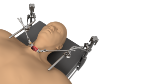
This is a lesion of a disc separating two vertebrae in your neck, which will be operated on from the front with the help of cloward cervical retractor. The scar will be on the front of your neck.
A little anatomical reminder: the human neck has seven cervical vertebrae all separated by shock absorbers: the intervertebral discs.
The lesion of the disc (disc disease), caused either by trauma or more often by wear and tear, can cause compression on nerve structures.
The cervical disc herniation sits at the back of the disc and can then compress either the nerve exiting between the two vertebrae (the nerve root) or even if the herniation is larger, compress the entire spinal cord which is located behind the disk.
When this compression acts on the outgoing nerve (root), it causes pain in the arm called cervico-brachial neuralgia, sometimes reaching the fingers. The painful path provides information on the level of the affected disc.
SPONTANEOUS EVOLUTION IN THE ABSENCE OF TREATMENT
The greater the compression and the longer the duration, the greater the risk of serious injury. The initial pain may then intensify, followed by tingling and loss of feeling or even problems with loss of strength in the hand or even the arm.
The pain is usually worsened by movement of the head and neck.
When the hernia is very large, and located in the middle of the disc, it can directly compress the main nervous structure, the spinal cord. This can result in a feeling of weakness in the legs (limiting your walking range) and dropping objects.
The spontaneous course can in some cases lead to an improvement or disappearance of neuralgia pain, but we can only consider waiting in the absence of deficient neurological disorders and if the pain remains bearable.
EXAMINATIONS TO BE MADE
Even if the surgeon examining you may have an already precise idea of the disc level causing the problem, a number of additional examinations are now useful to confirm the exact location of the lesion but also to eliminate a a number of diseases which could give misleading signs.
These are the following exams:
Standard radiography: it allows recognizing the level of the affected disc, because in general, the disc is less high and partially crushed. Most often, hernias are located between the 5th and 6th cervical vertebrae and / or between the 6th and 7th cervical vertebrae.
The x-ray will also look for signs of osteoarthritis in older lesions, and also study the general curvature of the cervical spine. This radiological examination does not present a danger of irradiation, except if it was repeated very often.
It is sometimes useful to order X-rays in the flexion and extension position to assess the flexibility of the spine and the instability of the affected disc.
The scanner (or tomodensitometry): which also uses X-rays, makes it possible to visualize the disc in a more precise way, by slices. It makes it possible to clearly specify the size of the hernia, its position and whether it is accompanied by bone constructions linked to osteoarthritis. Sometimes the radiologist decides to give an intravenous injection of iodine, so that they can better see the surroundings of the hernia. You should know that the scanner uses a much larger quantity of X-rays than the simple radiography.
Magnetic resonance imaging (MRI): it gives additional information to that of the scanner, by showing the surrounding soft tissues better and by providing information on the quality of the tissues, it shows on the other hand the structures less well. It is particularly good at detecting lesions of the spinal cord (cervical myelopathy) in cases of narrowing of the duct due to osteoarthritis or a very large hernia.
Electromyogram (EMG): It allows to study the quality of functioning of the nerves, to confirm and to specify the severity of the nerve compression.
More complex examinations (motor and sensory evoked potentials) are sometimes required in order to record the functioning of the spinal cord.
These examinations make it possible to take into account the severity of the nerve damage, which sometimes explains the differences in recovery.
For more details about surgical instruments, please visit: jimymedical.co.uk